Product Summary

Cat. No.
DIA 011-01
Specificity
DIA 011-01 binds both membrane-bound MAC (active) and fluid-phase SC5b-9 complexes (inactive). (1)
Immunogen
Purified C5b-9
Host
Mouse
Positive Species Reactivity
Human, Pig, Horse, Baboon
Negative Species Reactivity
Not determined
Isotype
IgG2a/k
Gene ID
727
Clone Number
aE11
Epitope Specificity
DIA 011-01 binds to a neoepitope exposed on C9
Unit Size
400 µL: Cat. No. DIA 011-01-04 1 mL: Cat. No. DIA 011-01-1 1 mg/mL +/- 15%. See Certificate of Analysis for details.
Purification
Protein A/G purified (from culture supernatant)
Form
Liquid
Solvent
0.01 M phosphate buffer, pH 7.4, containing 0.15 M NaCl, 15mM NaN3
Conjugation
Unconjugated BSA free
Storage
4-8ºC without exposure to light. No precautions necessary during handling.
Target
C5b-9 is also known as the terminal complement complex (TCC). The TCC consists of C5b, C6, C7, C8 and C9 and forms the membrane attack complex (MAC) as well as the non-lytic fluid-phase SC5b-9 complex (with protein S). The MAC forms channels in target cell membranes leading to cell lysis by osmotic leakage. The complexes contain neoantigens that are absent from the individual native components from which they are formed and DIA 011-01 is directed against a neoepitope exposed on C9 when incorporated into the TCC.
Other Info
This product is not for further manaufacture.

Application

ELISA: DIA 011-01 is well suited for quantifying TCC in ELISA. (2,3)

WB: DIA 011-01 is not recommended for Western blotting as the epitope is destroyed during the process. (4)

IHC: TCC in various tissues by immunohistochemistry (frozen and paraffin sections). (1, 2, 4)

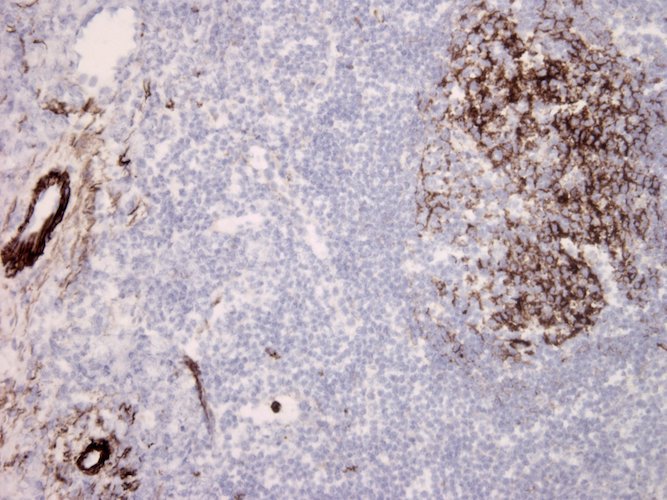
IHC staining of frozen tonsil section. Vascular endothelia and dendritic cells of germinal center have strong and intensive label. Dilution: 1:10

IF: DIA 011-01 can be used in immunofluorescence staining. (5)

Scientific References

1. Mollnes TE, Harboe M (1987) Immunohistochemical detection of the membrane and fluid-phase terminal complement complexes C5b-9(m) and SC5b-9. Consequences for interpretation and terminology. Scand J Immunol 26:381-6.

2. Mollnes TE, Lea T, Harboe M, Tschopp J (1985) Monoclonal antibodies recognizing a neoantigen of poly (C9) detect the human terminal complement complex in tissue and plasma. Scand J Immunol 22:183-195.

3. Palarasah,Y, Nielsen C, Sprogøe U, Christensen ML, Lillevang S, Madsen HO, Bygum A, Koch C, Skjodt K, Skjoed MO (2011) Novel assays to assess the functional capacity of the classical, the alternative and the lectin pathways of the complement system. Clin Exp Immun 164: 388-395.

4. Mollnes TE (1997) Analysis of in vivo complement activation. Herzenberg LA, Weir DM, Herzenberg LA, Blackwell C: Weir’s Handbook of Experimental Immunology. Boston, MA: Blackwell Science, pp. 78.1-78.8.

5. Jansen J, Høgåsen K, Mollnes T (1993) Extensive Complement Activation in Hereditary Porcine Membranoproliferative Glomerulonephritis Type 11 (Porcine Dense Deposit Disease. Am J Path 194: 1356-1365.

Cat. No.
DIA 011-01
Specificity
DIA 011-01 binds both membrane-bound MAC (active) and fluid-phase SC5b-9 complexes (inactive). (1)
Immunogen
Purified C5b-9
Host
Mouse
Positive Species Reactivity
Human, Pig, Horse, Baboon
Negative Species Reactivity
Not determined
Isotype
IgG2a/k
Gene ID
727
Clone Number
aE11
Epitope Specificity
DIA 011-01 binds to a neoepitope exposed on C9
Unit Size
400 µL: Cat. No. DIA 011-01-04 1 mL: Cat. No. DIA 011-01-1 1 mg/mL +/- 15%. See Certificate of Analysis for details.
Purification
Protein A/G purified (from culture supernatant)
Form
Liquid
Solvent
0.01 M phosphate buffer, pH 7.4, containing 0.15 M NaCl, 15mM NaN3
Conjugation
Unconjugated BSA free
Storage
4-8ºC without exposure to light. No precautions necessary during handling.
Target
C5b-9 is also known as the terminal complement complex (TCC). The TCC consists of C5b, C6, C7, C8 and C9 and forms the membrane attack complex (MAC) as well as the non-lytic fluid-phase SC5b-9 complex (with protein S). The MAC forms channels in target cell membranes leading to cell lysis by osmotic leakage. The complexes contain neoantigens that are absent from the individual native components from which they are formed and DIA 011-01 is directed against a neoepitope exposed on C9 when incorporated into the TCC.
Other Info
This product is not for further manaufacture.

ELISA: DIA 011-01 is well suited for quantifying TCC in ELISA. (2,3)

WB: DIA 011-01 is not recommended for Western blotting as the epitope is destroyed during the process. (4)

IHC: TCC in various tissues by immunohistochemistry (frozen and paraffin sections). (1, 2, 4)

IHC staining of frozen tonsil section. Vascular endothelia and dendritic cells of germinal center have strong and intensive label. Dilution: 1:10

IF: DIA 011-01 can be used in immunofluorescence staining. (5)

1. Mollnes TE, Harboe M (1987) Immunohistochemical detection of the membrane and fluid-phase terminal complement complexes C5b-9(m) and SC5b-9. Consequences for interpretation and terminology. Scand J Immunol 26:381-6.

2. Mollnes TE, Lea T, Harboe M, Tschopp J (1985) Monoclonal antibodies recognizing a neoantigen of poly (C9) detect the human terminal complement complex in tissue and plasma. Scand J Immunol 22:183-195.

3. Palarasah,Y, Nielsen C, Sprogøe U, Christensen ML, Lillevang S, Madsen HO, Bygum A, Koch C, Skjodt K, Skjoed MO (2011) Novel assays to assess the functional capacity of the classical, the alternative and the lectin pathways of the complement system. Clin Exp Immun 164: 388-395.

4. Mollnes TE (1997) Analysis of in vivo complement activation. Herzenberg LA, Weir DM, Herzenberg LA, Blackwell C: Weir’s Handbook of Experimental Immunology. Boston, MA: Blackwell Science, pp. 78.1-78.8.

5. Jansen J, Høgåsen K, Mollnes T (1993) Extensive Complement Activation in Hereditary Porcine Membranoproliferative Glomerulonephritis Type 11 (Porcine Dense Deposit Disease. Am J Path 194: 1356-1365.

×